Saturday, December 15, 2012

Migraine


Abortive Medication Stratification by Headache Severity
ModerateSevereExtremely Severe
NSAIDsNaratriptanDHE (IV)
IsomethepteneRizatriptanOpioids
ErgotamineSumatriptan (SC,NS)Dopamine antagonists
NaratriptanZolmitriptan
RizatriptanAlmotriptan
SumatriptanFrovatriptan
ZolmitriptanEletriptan
AlmotriptanDHE (NS/IM)
FrovatriptanErgotamine
EletriptanDopamine antagonists
Dopamine antagonists
Table 2. Preventive Drugs
First lineHigh efficacyBeta-blockers


Tricyclic antidepressants


Divalproex


Topiramate


Low efficacyVerapamil


NSAIDs


SSRIs


Second lineHigh efficacyMethysergide


Flunarizine


MAOIs


Unproven efficacyCyproheptadine


Gabapentin


Lamotrigine


Comorbid ConditionMedication
HypertensionBeta-blockers
AnginaBeta-blockers
StressBeta-blockers
DepressionTricyclic antidepressants, SSRIs
UnderweightTricyclic antidepressants
EpilepsyValproic acid, Topiramate
ManiaValproic acid

Sunday, December 9, 2012

Carcinoma Breast

In hormone receptor-positive, HER2-negative disease, endocrine therapy should be a clinician’s first choice. Single-agent chemotherapy and combination chemotherapy are also possible options. Chemotherapy and hormone therapy should not be given concomitantly.

For postmenopausal women with ER-positive:
Table 1. Available Endocrine Therapies
Agent ClassDrugs Available
Anabolic steroidsnandrolone decanoate
AIs, third-generation, nonsteroidalanastrozole; letrozole
AIs, third-generation, steroidalexemestane
Estrogensestrogens
ER downregulatorfulvestrant
Luteinizing hormone-releasing hormone analoguesgoserelin; leuprorelin; triptorelin
Progestinsmedroxyprogesterone acetate; megestrol acetate
Selective ER modulatorstamoxifen, toremifene

Letrozole- oral non steroidal aromatase inhibitor 
For postmenopausal women with ER-positive disease, several endocrine therapy options are considered standard. If AIs have not been used in the adjuvant setting, several studies have shown they are superior to other endocrine therapy, including tamoxifen. The third-generation AIs — letrozole, exemestane, and anastrozole — provided statistically significant survival benefits. Fulvestrant, an ER downregulator, is available in 2 doses: 250 mg or 500 mg. Recently published data from the phase 2 FIRST trial compared fulvestrant with anastrozole as first-line endocrine therapy for postmenopausal women with hormone receptor-positive advanced breast cancer. Specifically, they compared high-dose fulvestrant (500 mg intramuscularly on days 0, 14, and 28, and monthly thereafter) with anastrozole (1 mg per day). Median time to progression (TTP), the primary endpoint, was 23.4 months in the fulvestrant arm and 13.1 months in the anastrozole arm (HR, 0.66; 95% CI, 0.47-0.92; = .01).


Bone-modifying agents beneficial in bone secondaries of breast CA (also caused by hormonal therapy):
1 of 3 bone-modifying agents:

Denosumab-targets RANKL(RANK ligand)-a protein that acts as the primary signal for bone removal. In many bone loss conditions, RANKL overwhelms the body's natural defenses against bone destruction.-120 mg subcutaneously every 4 weeks,
intravenous (IV) pamidronate (90 mg over no less than 2 hours),
 or zoledronic acid (4 mg over no less than 15 minutes every 3 to 4 weeks)

Other Agents:
Everolimus- inhibitor of Mammalian Target of Rapamycin (mTOR)- an immunosuppressant
Patients who become resistant to endocrine therapy have a poor prognosis. The targeted agent everolimus, which has just been approved, helps patients circumvent endocrine resistance. Resistance to endocrine therapy has been associated with the activation of the P13K-mTOR pathway, which, in turn, activates the ER. Everolimus inhibits the mTOR pathway.

Bevacizumab-monoclonal antibody-angiogenesis inhibitor-inhibits VEGF-A. The use of bevacizumab in breast cancer has been surrounded by controversy in recent years. In 2009, the EMA approved the combination of paclitaxel and bevacizumab in MBC, based on the E2100 clinical trial. This study showed a 5.5-month improvement in PFS for the combination over paclitaxel monotherapy. With longer follow-up of E2100, however, investigators found the PFS benefit did not translate into an OS benefit.


HER2-Positive Metastatic Breast Cancer

Trustuzumab- Monoclonal antibody that interfers with HER2/nu receptor

More recently, level 1 evidence has shown that clinicians should continue blockade of the HER2 pathway even after a patient progresses on an anti-HER2 agent. The German Breast Group enrolled 78 patients with HER2-positive breast cancer that had progressed during treatment with trastuzumab and randomized them to received capecitabine alone or in combination with trastuzumab.
Lapatinib-Dual tyrosine kinase inhibitor- Inhibitor of  HER2/nu receptor + EGFR pathways

ESMO guidelines state that available evidence suggests continuing anti-HER2 therapy for as long as possible.

Capecitabine:
5-FU prodrug, converted enzymatically to 5-FU in tumour

Pertuzumab:
The first of its class in a line of agents called "HER dimerization inhibitors"

Table 2. Available Chemotherapy Agents
Anthracycline-containing regimensDoxorubicin or epirubicin monotherapy
Doxorubicin/cyclophosphamide or epirubicin/cyclophosphamide
Liposomal doxorubicin with or without cyclophosphamide
Fluorouracil/doxorubicin/cyclophosphamide or fluorouracil/epirubicin/cyclophosphamide
Taxane-containing regimensPaclitaxel monotherapy
Docetaxel monotherapy Nab-paclitaxel
Anthracycline (doxorubicin or epirubicin)/taxane (paclitaxel or docetaxel)
Docetaxel/capecitabine
Paclitaxel/gemcitabine
Paclitaxel/vinorelbine
Paclitaxel/carboplatin
Newer cytotoxic regimensEribulin
Ixabepilone (not approved by EMA)
Cyclophosphamide/methotrexate/fluorouracil (CMF)
Platinum-based combinations (eg, cisplatin + 5-fluouracil; carboplatin + gemcitabine)
Capecitabine
Vinorelbine
Capecitabine + vinorelbine
Vinorelbine with or without gemcitabine
Oral cyclophosphamide with or without methotrexate


Eribulin

The recently approved chemotherapeutic eribulin mesylate is only approved for heavily pretreated women with locally recurrent or MBC. The label states it is approved for patients who have progressed after at least 2 chemotherapeutic regimens for advanced disease.

Brain Metastasis:
Brain metastases occur in 30% to 50% of patients with HER2-positive MBC. Patients with a small number of potentially resectable brain metastases should be treated with surgery or radiosurgery. Patients who respond to anti-HER2-based therapy and have controlled extracranial disease can live for several years after the diagnosis and treatment of brain metastases. Recently the phase 2 LANDSCAPE trial, investigating the combination of lapatinib and capecitabine for the treatment of previously untreated brain metastases from HER2-positive MBC, showed that 65.9% had an objective central nervous system (CNS) response.
Results from a phase 2 trial have shown that brain metastases can respond to a combination of lapatinib plus capecitabine

Rebiopsy:
National Comprehensive Cancer Network (NCCN), and others state that hormone receptor status and HER2 status should be re-evaluated at least once in a metastatic lesion.

NOTE- These are personal notes.They don't qualify be quoted.

Saturday, December 8, 2012

Pericardium


The heart and its pericardium make up the contents of the middle mediastinum. The left and right phrenic nerves and their adjacent arteries (pericardiacophrenic) lie to the left and right of the pericardium and anterior to the roots of the lungs.

Parietal and visceral pericardium are continuous. This continuity takes place at the points where the major blood vessels enter and leave the heart. The parietal pericardium has two inseparable parts, an outer fibrous part and an inner smooth part, the serous part.The potential space between the visceral and serous parietal pericardium is the pericardial cavity.

Second View:


The fibrous pericardium is the outermost layer, and it is firmly bound to the central tendon of the diaphragm. Extrapericardial fat, which may be visible radiographically, is often found in the angles between the pericardium and diaphragm on each side. The pericardium is attached to the sternum (by the sternopericardial ligaments) and is adherent to the mediastinal pleura except where the two are separated by the phrenic nerves.
The serous pericardium  is a closed sac, the parietal layer of which lines the inner surface of the fibrous pericardium and is reflected onto the heart as the visceral layer, or epicardium. The potential space between the parietal and visceral layers contains a thin film of fluid and is known as the pericardial cavity.


Pericardial Sinuses


Within the pericardial cavity, at the points where the visceral and parietal pericardia are continuous with one another, small chambers or sinuses are located. In this diagram, the heart has been removed and you are looking toward the posterior wall of the pericardial cavity.


The pericardial sinuses:
  • transverse pericardial sinus
  • oblique pericardial sinus
The transverse pericardial sinus can easily be reached by sticking your finger between the superior vena cava and the ascending aorta and pulmonary trunk. This sinus is a leftover from heart development in the embryo.
Again, slide two or three fingers under and behind the heart until they reach a dead end. Your fingers are now in the oblique pericardial sinus.

1. The pericardium starts to form during the _______ week of developme

Correct
. 5th

2. Which statement is true about the pleuropericardial folds?


Correct answer:
a. they partition the thorax into a pericardial cavity and two pleural cavities

3. What anatomic structure is located within, and migrates with, each pleuropericardial fold?


Correct answer:
b. phrenic nerve

4. The pleuropericardial folds initially form and grow along a _________ plane.


Correct answer:
d. coronal (frontal)


Sunday, September 16, 2012

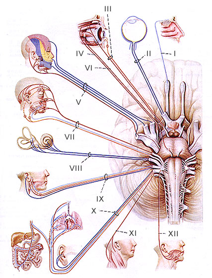
Cranial Nerves


CN which is the smallest -- olfactory n.
CN which enters cerebrum directly - olfactory nerve
CN with longest intracranial (subarachnoid) course -- trochlear n
CN which emerges posterior to brain stem- trochlear nerve
CN with dorsal exit -- trochlear n.
CN which is the largest and thickest -- trigeminal n.

CN which is largest -- trigeminal nerve
CN with longest extracranial course --vagus n.
CN having longest intraosseous course -- facial nerve
CN with longest  intradural course - abducent nerve
CN passing through cavernous sinus -- abducent nerve
CN involved in raised intracranial tension -- abducent nerve 
Abducent nerve has the longest intra-cranial INTRADURAL course!!
Thickest nerve is SCIATIC nerve
Thickest cutaneous nerve is GREATER OCCIPITAL nerve
Labourer’s nerve-median nerve
Dentist’s nerve-inferior alveolar nerve
Alderman’s nerve-auricular branch of vagus nerve
Nerve of laterjet-largest gastric branch of vagus nerve


CN with longest intracranial course -- trochlear n.
CN with longest extracranial course --vagus-

Longest nerve:CN X (Vagus) which reaches from the medulla to the digestive and urinary organs   
CN which is the largest and thickest -- trigeminal n.
CN with dorsal exit -- trochlear n.
CN which is the smallest -- olfactory n.


Longest course within the skull is the trochlear, while Largest is trigeminal,
longest outside skull is vagus-from medulla to digestive and urinary organs

Saturday, July 21, 2012

Thyroid Replacement Therapy


Complications of overreplacement with levothyroxine sodium Include the following:
  • Accelerated bone loss
  • Reduction in bone mineral density
  • Osteoporosis
  • Increased heart rate
  • Increased cardiac wall thickness
  • Increased contractility
The last three problems above increase the risk of cardiac arrhythmias (especially atrial fibrillation), particularly in the elderly population.

Medications that suppress TSH production include steroids, dopamine, dobutamine, and octreotide.


Up to 30-40% of patients with hypothyroidism have anemia, usually from decreased erythropoiesis. In 15% of patients, the anemia is of the iron deficiency type, with microcytosis and hypochromia. Although this can be a normocytic normochromic anemia, the most common morphologic abnormality is a macrocytic anemia that may be partially due to insufficient vitamin B-12 and folate intake.


Glomerular filtration rate, renal plasma flow, and renal free water clearance are all decreased in hypothyroidism and may result in hyponatremia.


Prolactin may be elevated in primary hypothyroidism. This is thought to be caused by overlap secretion due to stimulation of the lactotroph by the elevated TRH level. The decreased clearance of prolactin in hypothyroidism may also play a contributory role. The elevated prolactin level leads to decreased gonadotropin secretion and decreased responsiveness to GnRH. The result of this is anovulatory cycles with menstrual abnormalities, galactorrhea, and infertility in some patients.


Other studies may be performed in the evaluation of complications of primary hypothyroidism (when indicated). These tests are usually not performed and are not necessary in routine diagnosis or evaluation of hypothyroid patients.
  • Chest radiograph - May show small pleural effusions
  • Electrocardiogram (ECG) - May show low-voltage QRS tracing, nonspecific ST-wave changes, and premature ventricular contractions; prolongation of the QT interval with torsade de pointes and ventricular tachycardia may be noted
  • Echocardiogram - May show some pericardial effusion in severe cases of hypothyroidism


    Long-Term Monitoring

    Upon the initiation of the levothyroxine replacement therapy, check thyroid function tests, specifically TSH, initially every 6-8 weeks as dose adjustments are made. After the attainment of the clinical euthyroid state and a normal TSH level, patients and the TSH levels may be checked every 6-12 months.
    More frequent follow-up and TSH checks may need to be performed when patients start taking medications, such as ferrous sulphate, calcium supplementation, and multivitamins, that have the potential to impair the absorption of levothyroxine and therefore to affect the TSH level. Patients need to be advised to separate these medications from levothyroxine by at least 4 hours.
    Follow-up care should include clinical evaluation for symptoms of hypothyroidism or iatrogenic hyperthyroidism.
    Physical examination should routinely include weight measurement, pulse and blood pressure determinations, and thyroid examination for the presence of nodules.
    Yearly thyroid ultrasonographic evaluation is important in patients with Hashimoto thyroiditis because of the increased risk of thyroid nodules in these patients and for follow-up of patients with existing benign thyroid nodules.

Tuesday, April 10, 2012

Facies

Hippocratic face (also known as "Hippocratic facies"; eyes are sunken, temples collapsed, nose is pinched with crusts on the lips and the forehead is clammy)
moon face (also known as "Cushingoid facies")
elfin facies - Williams syndrome
Potter facies - oligohydramnios
mask like facies - parkinsonism
Leonine facies - lepromatous leprosy
Mitral facies - mitral stenosis
Amiodarone facies (deep blue discoloration around malar area and nose)
Acromegalic facies - acromegaly
flat facies - down's syndrome
Marfanoid facies - marfan's syndrome
snarling facies - myasthenia gravis
Myotonic facies - myotonic dystrophy
torpid facies - myxoedema
mouse facies - chronic renal failure
plethoric facies - cushing's syndrome and polycythemia vera
'bird-like' facies- pierre robin sequence
ashen grey facies - myocardial infarction
gargoyle facies - hurler's syndrome
monkey facies - marasmus
hatchet facies - myotonia atrophica
gorilla-like face - acromegaly
bovine facies or cow face - craniofacial dysostosis or crouzons syndrome
marshall halls facies - hydrocephalus
frog face - intra nasal disease

Friday, March 23, 2012

Fractures

break in continuity of bone
  • Traumatic-sustained due to excessive force-a qualified fracture usually means traumatic fracture
  • Pathological-usually trivial-bone already weak by underlying disease
  • Displaced fracture may occur due to fracturing force/muscle pull on fracture filaments/gravity-displacement may be shift/angulation/rotation
  • Simple/closed
  • Compound/open-
  1. internal compounding-piercing of sharp fracture end
  2. external compounding-lacreation+fracture from outside

  • Transverse # fracture line perpendicular to long axis of bone, caused by tapping/bending force
  • Oblique #line of # oblique, caused by a bending force which in addition has component along the long axis of the bone
  • Comminuted #with miltiple fragments,caused by crushing/compression force along the long axis of the bone
  • Segmental # two fractures in same bone@different levels

Fractures with Eponyms
  • Monteggia #dislocation #proximal 1/3rd of ulna + dislocation of head of radius
  • Galeazzi #dislocation # distal 1/3rd of radius with dislocation of distal radio-ulnar joint
  • Night stick # isolated # shaft of ulna
  • Colles' # occuring in adults@cortico-cancellous junction of distal end of radius + dorsal tilt & other displacements
  • Smith's # occur in adults@cortico-cancellous junction of distal end of radius with ventral tilt & other displacements (reverse Colles')
  • Barton's (marginal) # intra-articular# through distal articular surface of radius, taking a margin, anterior/posterior of the distal radius with the carpals displaced anteriorly/posteriorly
  • Chauffeur's # intra-articular oblique # of styloid process of radius
  • Bennet's # dislocation-oblique,intra-articular # of base of 1st metacarpal with sublaxation of trapezio-metacarpal joint
  • Rolando # extra articular # of base of 1st metacarpal
  • Boxer's # a ventrally displaced # through the neck of the 5th metacarpal-common in boxers
  • Side swipe # an elbow injury, combination of #distal end of humerus with # proximal end of radius &/or ulna (baby car #)
  • Bumper # comminuted depressed type-lateral condyle of tibia
  • Pott's # bimalleolar ankle
  • Cotton's # trimalleolar ankle
  • Massonaise's # ankle with # neck of fibula
  • Pilon # comminuted, depressed intra-articular # distal end of tibia
  • Aviator's # neck of talus
  • Chopart # dislocation # dislocation through inter tarsal joints
  • Jone's # Avulsion # base of 5th metatarsal
  • Jeffersen's # 1st cervical vertebra
  • Whiplash Injury- cervical spine injury involving sudden flexion followed by hyperextension
  • Chance # also called seat belt #, line of # runs horizontally through the body of vertebra through & through to the posterior elements
  • March # fatigue # of shaft of 2nd & 3rd metatarsal
  • Burst # comminuted # of vertebral body where fragments burst out in different directions
  • Clay-Shoveller's # an avulsion # of spinous process of one or more of the lower cervical or upper thoracic vertebra
  • Hangman's # pedicle & lalamina of C2 vertebra, with sublaxation of C2 over C3-sustained in hanging
  • Dashboard # posterior lip # of acetabulum, often associated with posterior dislocation of hip
  • Straddle # bilateral superior & inferior pubic rami #
  • Malgaigne's # a type of pelvis # in which there is a combination of #s- pubic rami anteriorly & SI joint/ilium posteriorly, on the same side
  • Mallet Finger- a finger flexed at the DIP joint d/t avulsion or rupture of extensor tendon@base of distal phallanx

Skin: Histology

Continually renewing stratified squamous epithelium-keratinizes & gives rise to derivative structures called appendages (pilosebaceous units, nails & sweat glands)

Cell Cycle-G0-G1-S-G2-M

Keratinocyte-ectodermally derived cell constituting 80% of epidermal cells-keratin filaments are hallmarks of keratinocytes & other epithelial cells

Immigrant cells- Melanocytes& Langerhans cells(migrate into epidermis during embryonic development), Merkel cells(differentiate in situ)

Melanocytes- located in stratum basale(stratum germinativum)- project their dendrites into epidermis- transfer their melanosomes (melanin packed membrane bound organelles) to keratinocytes which contains majority of melanin pigment - 36 keratinocytes per melanocytes- epidermal melanin unit


Keratinization- genetically programmed, carefully regulated, complex series of morphological changes & metabolic events that occur progressively in postmitotic keratinocytes & involve-

  • increased cell size & flattening
  • appearance of new cellular organelles & structural reorganization of those present
  • change from generalized cellular metabolism to a more focussed metabolism associated with the synthesis of molecules related to keratinization(structural proteins & lipids)
  • alterations in properties of plasma membrane, cell surface antigen & receptors
  • eventual degradation of cellular organelles including internucleosomal chromatin fragmentation characteristic of apoptosis
  • dehydration

Epidermal Layers
  • Basal layer/stratum germinativum-attached to basement membrane-contains mitotically active keratinocytes-contains house keeping organelles(RER,golgi complex, mitochondria, lysosomes & ribosomes)-gives rise to superficial layer
  • Spinous layer/Stratum spinosum-named for spine like appearance of cell margins in histological sections. Spines are abundant desmosomes, calcium dependent cell surface modifications that promote adhesion of epidermal cells & resistance to mechanical stress.Upper spinous layer cells have organelles called lamellar granules
  • Malpighian layer-includes both basal & spinous layer
  • Granular layer/stratum granulosum-characterised by buildup of components necessary for the process of programmed cell death & formation of a superficial water impermeable layer.The most apparent structures within these cells are-basophilic keratinohyaline granules-composed of profilaggrin,keratin intermediate filament & loricrin. Conversion of profilaggrin to filaggrin(filament aggregating protein)-occurs during transition of granular to cornified cells
  • Corny layers or Stratum corneum- formed of cornified or horny cells which is the largest of epidermis & have highest concentration of free amino acids,esp in mid layers.Stratum corneum cells retain some metabolic function (not just an inert covering)
Normal turnover time of epidermis/skin doubling time-- 4wks
VLBW/premature infants lack-stratum corneum

Sunday, March 18, 2012

Carpal Tunnel

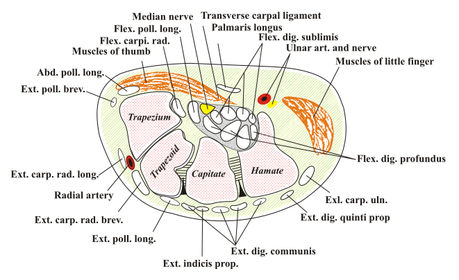
  • Carpal tunnel or carpal canal is the passageway on the palmar side of the wrist that connects the forearm to the middle compartment of the deep plane of the palm

  • The canal is narrow and when any of the nine long flexor tendons passing through it swells or degenerates, the narrowing of the canal often results in the median nerve becoming entrapped or compressed, a medical condition known as carpal tunnel syndrome

  • The structures passing through it are
  1. flexor digitorum profundus (four tendons)
  2. flexor digitorum superficialis (four tendons)
  3. flexor pollicis longus (one tendon)
A single nerve passes through the tunnel: the median nerve between tendons of flexor digitorum profundus and flexor digitorum superficialis

The carpus, the bony elements of the wrist, form an arch which is convex on the dorsal side of the hand and concave on the palmar side. The groove on the palmar side, the sulcus carpi, is covered by the flexor retinaculum, a sheath of tough connective tissue, thus forming the carpal tunnel. The flexor retinaculum is attached radially to the scaphoid tubercle and the ridge of trapezium, and on the ulna side to the pisiform and hook of hammate

The narrowest section of the tunnel is located a centimetre beyond the mid-line of the distal row of carpal bones where the sectional area is limited to 1.6 cm2

The tendons of the flexor digitorum superficialis and profundus pass through a common ulnarsheath, while the tendon of the flexor pollicis longus passes through a separate radial sheath. Themesotendon shared by these tendons is attached to the radial and palmar walls of the carpal tunnel

Superficial to the carpal tunnel and the flexor retinaculum, the ulnar artery and ulnar nerve pass through the ulnar tunnel

Movements in the wrist affects the shape and width of the carpal tunnel. The width decreases considerably during normal range of motion in the wrist and because the carpal bones move in relation to each other with every motion of the hand the bony walls of the tunnel are not rigid. Both flexion and extension increase compression in the carpal tunnel:

  • Flexing the wrist causes the flexor retinaculum to move closer to the radius which considerably decreases the cross section of the proximal opening of the tunnel. Additionally, the distal end of the capitate presses into the opening.
  • In extreme extension. the lunate constricts the passage as it is pressed toward the interior of the tunnel.


File:Carpal-Tunnel.svg

Thursday, March 15, 2012

Biosimilar Insulins

Biocon is conducting Phase-3 trials of recombinant human insulin (biosimilar of Novo Nordisk’s Novolog) in Europe and global Phase-1 trials for glargine (biosimilar of Sanofi-Aventis’ Lantus). Biocon will continue to develop other biosimilars — aspart (biosimilar of Novo Nordisk’s Novolog) and lispro (biosimilar of Eli Lilly’s Humulog)

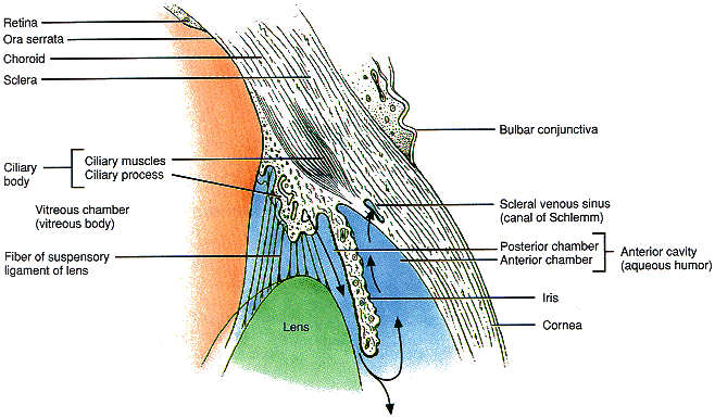
Eye: Anatomy


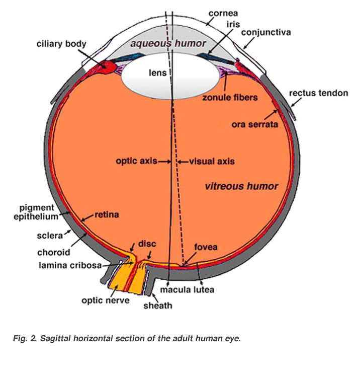
Outer Layer- cornea/sclera


anterior part of sclera covered by mcous menbrane-the conjunctiva

Cornea:
  • 3 layers-epithelium, substantia propria(stroma),Descemet's membrane with endothelium
  • epithelium-stratified, basal cells lie on Bowman's membrane
  • stroma-90% of corneal thickness - regularly arranged thin fibrils of collagen ensheathed by acid mucopolysaccharides set in a ground substance- form ribbon like bundles & give the stroma a laminated appearance- fibrils circular in CS-spaced equidistant- hexagonal lattice
  • transparency related to regularity of stromal components-interfibrilar spacing less than a wavelength of light-tangential rows of fibres act as diffraction gratting resulting in destructive interference of scattered rays
  • Descemet's membrane-thin elastic membrane-covered on posterior surface by endothelium
  • stromal hydration maintained by endothelium- electrolytes removed & water flows passively
  • endothelium examined by 500x specular microscope
  • endothelial cells decrease in number with age-residual individual cells enlarge to compensate
  • corneoscleral junction- limbus- cornea set into a sclera like a watch glass
  • nerve supply-trigeminal
  • no blood vessels- minute arcades @ limbus (1mm)
  • corneal nourishment- diffusion of aqueous humour & peripheral vessels
Lining the Inner Aspect of Sclera-
  1. Uveal Tract-highly vascular-for nutrition
  2. Retina

Uveal Tract:
  • Choroid
  • Ciliary Body
  • Iris- anteriormost
Anterior Chamber:
  • aquous humour
  • between cornea & iris 2.5 mm deep in centre
  • peripheral recess- angle of anterior chamber
  • canal of Schlemm- circular venous sinus in inner layer of sclera- often more than one lumen
  • trabecular meshwork between canal of Schlemm & recess of anterior chamber
Iris:
  • anterior surface-single layer endothelium-not continuous@crypts
  • stroma contains branched connective tissue cells
  • iris stroma usually pigmented-unpigmented in blue sclera
  • blood vessels in iris-radial
  • tissue spaces communicate directly with anterior chamber through crypts@ ciliary border
  • thinnest@attachment to ciliary body
  • posterior surface-2 pigmented epithelium-developmentally from retina-continuous@pupillary margin-anterior layer flattened cells-posterior layer cuboidal cells
  • pupillary smooth muscles derived from from anterior epithelial cells
  • sensory- trigeminal
  • sphincter pupillae-occulomotor
  • dilator pupillae-sympathetic (cervical chain)
Ciliary Body:
  • like isosceles triangle- base forwards
  • chief mass-ciliary muscle-unstriped-3 parts-circumferential-blends with scleral spur
  • most muscles meridional in anteroposterior direction, 2nd portion v-shaped interdigitating concentrically@base of iris, 3rd portion -insertion@ root of iris-just anterior to pigmentary epithelium-closely related to dilator muscle
  • anterior surface-corrugated-pars plicata-contains ciliary processes(tufts of blood vessels-like glomeruli) in between
  • posterior part-smooth-pars plana
  • covered on inner surface by 2 layers of epithelium-belongs to retina-only outer layer pigmented
  • posterior extent-ora serrata-transition from ciliary body to choroid gradual
  • ora serrata more anterior on nasal side than temporal
  • sensory-trigeminal
  • motor-occulomotor & sympathetic
Choroid:
  • extremely vascular membrane in contact everywhere with sclera with a potential space-epichoroidal/suprachoroidal space in between
  • inner side lamina vitrea/membrane of Bruch
  • blood vessels increase in calibre from inside to outside-choriocapillaries(fenestrated vessels) immediately beneath membrane of Bruch
  • sensory-trigeminal
  • autonomic supply-for vasomotor
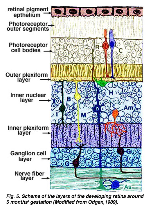
Retina:
  • Outer layer epithelium-hexagonal single layer pigment epithelium
  • Inner layer epithelium-suddenly changes@ora serrata into highly complex visual retina
Retina formed by 3 strata of cells & their synapses:
  • visual cells-externally
  • relay layer of bipolar cells-intermediate
  • ganglion cells-internally

  1. pigment epithelium-hexagonal cells-single layer-assist metabolism of retina- products of metabolism are freely exchanged between receptor cells & pigment epithelium-melanin granules prominent(absorbs light)-phagosomes present-
  2. rods & cones-neural epithelium-discs renewed continuously-rod discs have limited life,eventually lost to pigment epithelium
  3. external limiting membrane-perforated by rods & cones
  4. outer nuclear layer-nuclei of rods & cones
  5. outer plexiform layer-synaptic layer-transmissive region
  6. inner nuclear layer-nuclei of bipolar cells-
  7. inner plexiform layer-synaptic
  8. ganglion cell layer
  9. nerve fibre layer-axons of ganglion cells running centrally to optic nerve
  10. internal limiting membrane-separates retina from vitreous


  • Fibres of Muller-better developed vertical cells-supportive neuroglial cell- nutritive function

  • Fovea Centralis- at posterior pole (3mm in the temporal side of the optic disc)-only cones present- other layers almost completely absent- most sensitive part of retina

  • Macula lutea(yellow spot)-surrounds fovea centralis-nuclear layers get thinned out-plexiform layer present-ganglion cells heaped up into several layer(in stead of consisting of a single row of cells)-no blood vessels present-nourishment entirely by choroid)-more sensitive than other parts of retina,less than fovea

  • Optic Disc-fibres of nerve fibre layer pass into the optic nerve-other layers of retina stop abruptly@ the edge of the aperture in the scleral canal- spanned by transverse layer of connective tissue containing much elastic tissue(lamina cribrosa)-through its meshes the optic nerve fibres pass-on posterior side the nerve fibres abruptly become surrounded by medullary sheaths
Lens:

Biconcave mass of peculiarly differentiated epithelium-developed from invagination of surface ectoderm of the fetus(compare with plantar corns)-original surface goes inside@centre-peripheral cells correspond to the basal cells of epidermis-inner old cells undergo sclerosis-changes analogous to that of stratum granulosum in epidermis-becomes massed together in the form of nucleus-Lens is devoid of nerve supply


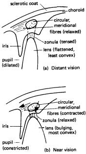
  1. Accommodation is one of the focusing mechanisms of the eye.
    Accommodation
  2. In order to focus rays from objects at varying distances, the lens must change it's refractive power.
  3. To change it's refractive power, the lens changes shape
  4. The exact shape of the lens is determined by seventy or so suspensory ligaments.
  5. The suspensory ligaments attached to the lens are called zonula.
  6. The zonula are attached radially around the lens.
  7. The zonula pull the edges of the lens towards the clilary body.
  8. When the eye is accommodated for distant vision, both the circular and meridional fibres of the ciliary muscle are relaxed. (a)
  9. When both the circular and meridional fibres of the ciliary muscle are relaxed, the zonula is stretched.
  10. When the zonula is stretched it pulls the elastic lens into a flattened shape.
  11. When the eye is accommodated for near vision, both the circular and meridional fibres of the ciliary muscle contract. (b)
  12. When both the circular and meridional fibres of the ciliary muscle contract, the tension in the zonula is released.
  13. When the ciliary muscle contracts, the ciliary process and choroid move forward toward the lens.
  14. When the tension in the zonula is released, it allows the the elastic lens to bulge.
  15. The lens of the eye is elastic.
  16. Because the lens is elastic, it bulges, shortens, and thickens.
  17. The ciliary process is the aqueous humor factory.
  18. The aqueous humour is drained out of the scleral venous sinus.


Tuesday, March 13, 2012

Sound Dynamics & Tests of Hearing

Decible Levels

Whisper 30 dB
Normal Conversation 60 dB
Shout 90 dB
Discomfort in ear 120dB
Pain in ear 130dB

Pitch is sensation of a particular frequency, dB is that of intensity

stapedial reflex is elicted with a sound of 70-100 dB
normal person can hear 20-20000 Hz
audiometric testing done 125-8000Hz

white noise-all frequencies if sound-comparable to white light

Narrow band noise-used in masking-it's a white noise without certain frequencies (above & below some patricular frequency having been filtered out)-used in pure tone audiometry

speech noise-noise having frequencies in speech range(300-3000 Hz)-all other frequencies filtered out

Masking-essential for all bone conduction tests-for air conduction test is required only when the difference in hearing between the two ears exceeds 40dB

Clinical Tests of Hearing

1.Finger Friction Test
2.Watch Test-practically obsolete
3.Speech (Voice Test)-conversational/forced whisper (use of spondee words by examiner)
4.Tuning Fork Tests- tests AC/BC

a)-Rinne Test (AC>BC is normal-positive Rinne)
BC>AC negative Rinne-minimum air-bone gap of 15-20 dB
  • Rinne test equal or negative for 256 Hz but positive for 512 Hz=> air bone gap 20-30 dB
  • Rinne negative for 256 & 512 Hz but positive for 1024 Hz => air bone gap 30-45 dB
  • Rinne negative for 256, 512 & 1024 Hz => air bone gap of 45-60 dB
Negative Rinne for 256, 512 & 1024 Hz indicates minimum AB gap of 15,30&45 dB respectively

False negative Rinne-in severe unilateral SNHL-correct diagnosis made by masking non test ear Barany's noise Box while testing for bone conduction-Weber lateralzation also helps

b)-Weber Test-done with 512 Hz (detects 15-20 dB)
  • lateralized to worse ear in conductive deafness
  • to better ear in sensorineural hearing loss
c)-ABC- cochlear function- comparison with examiner
d)-Schwabach's-ABC with an occluded meatus (reduced in SNHL, lengthened in CHL)

e)-Bing Test- effect of occlusion of meatus
  • Bing Positive-normal person/SNHL hear louder with occlusion of meatus
  • Bing Negative-CHL appreciates no change

f)- Gelle's Test- test of bone conduction, examined is the effect of increased air pressure in ear canal on hearing-has been replaced by tympanometry to find out stapes fixation
  • Positive in normal persons & SNHL-decreased hearing on increased pressure
  • Negative in fixed/disconnected ossicular chain

Audiometric Tests

1. Pure Tone Audiometry:
-Measure of cochlear function
-Air Conduction Threshold measured for tones 125,250,500,1000,2000,4000 & 8000 Hz
-Bone conduction threshold measured for 250,500,1000,2000 & 4000 Hz
-AB gap measured (normal individual has no AB gap in audiometry)

Shadow Curve- obtained from non-test better ear when difference between two ears is 40dB or more above air conduction thresholds-Masking is required if difference is>40dB(done by employing narrow band noise to the non-test ear)

2.Speech Audiometry:
-patient's ability to hear & understand speech is measured.parameters studied are
  • Speech Reception Threshold-normally SRT is within 10 dB of the average pure tone threshold of 3 speech frequencies(500,1000 & 2000 dB).SRT better than pure tone average by more than 10 dB suggests a functional hearing loss

  • Discrimination Score(DS) or Speech Recognition Score (SRS)
Normal 90-100%
Slight Difficulty 76-88%
Moderate difficulty 60-74%
Poor 40-58%
Very Poor <40%


Optimum Discrimination Score (ODS)-expressed in %
Half Peak Level (HPL) expressed as dB-a derived figure from audiogram

  • Normal- ODS 100% @30 dB,HPL-15 dB
  • Conductive Deafness of 40 dB-ODS100% @70 dB,HPL-55 dB
  • SNHL of 40 dB-ODS never reaches 100%, constant beyond a level
  • Retrocochlear loss- Roll over curve obtained, score falls below ODS beyond a frequency
3.Bekesy Audiometry-no longer used
4.Tympanometry-220 Hz tone delivered

  • Type A-normal tympanogram
  • As-fixation of sosicles-otosclerosis/malleus fixation-lower compliance@ambient air pressure
  • Ad-ossicular discontinuity/thin & lax TM -high compliance at or near ambient pressure
  • Type B-flat or dome shaped graph- no change in compliance with pressure changes-seen in middle ear fluid or thick TM
  • Type C-maximum compliance occurs with negative pressures in excess of 100 mm H2O-seen in retracted TM & may show some fluid in middle ear
Acoustic Reflex: Presence of stapedial reflex @ lower intensities (40-60 dB) than usual 70-100dB indicates recruitment & thus a cochlear type of hearing loss

Stapedial Reflex Decay- VIII th nerve lesion - if a sustained tone of 500-1000 Hz delivered 10dB above the acoustic reflex threshold,for a period of 10 seconds, brings the reflex amplitude to 50%, shows abnormal adaptation

Absence of stapedial reflex when hearing is normal indicates lesion of facial nerve, proximal to the nerve to stapedius.

Special Tests of Hearing


  1. Recruitment:
  • phenomenon of abnormal growth of loudness
  • the ear which doesn't hear low intensity sounds begins to hear greater intensity sounds as loud or even louder than normal hearing ear
  • poor candidates of hearing aid
  • cochlear lesions (Menier's Disease,presbycusis)
  • Alternate binaural loudness balance test- used to detect recruitment in unilateral cases
2. Short Increment Sensitivity Index
  • Patients with cochlear lesions distinguish smaller changes in intensity of pure tone better than normal persons & those with CHL or retrocochlear lesions
  • CHL- SISI score<15%
  • Cochlear Lesion-SISI score 70-100%
  • Nerve Deafness- SISI score 0-20%
3. Threshold Tone Decay Test:
  • measure of nerve fatigue
  • normal person can hear a tone continuously for 60 seconds, nerve fatigue-it's less
  • result expressed as dB decay
  • decay> 25dB=> retrocochlear lesion
4. Evoked Response Audiometry:
  • Electrocochleography (EcoG)-measures electrical potentials arising in the cochlea & CN VIII in response to auditory stimuli within 5 mili seconds, the response is in the form of cochlear microphonics,summating potentials & action potentials of VIII th nerve-finds threshold of hearing in young infants & children to within 5-10 dB
  • Auditory Brain Stem Responses-non-invasive- ECOLI,MA-7 wave forms I to VII-wates are studied for absolute latency, inter wave latency, amplitude
5.Otoacoustic Emissions: produced by outer hair cells of cochlea
6.Central Auditory Tests:designed to find defects in central auditory pathways & temporal cortex
7. Hearing Assessment ininfants & Children:
  • Screening procedures- arousal test & auditory response cradle
  • Behaviour Observation Audiometry- Moro's Reflex, Cochleo Palpebral Reflex & Cessation Reflex
  • Distraction techniques
  • Coonditioning Techniques
  • Objective Tests