


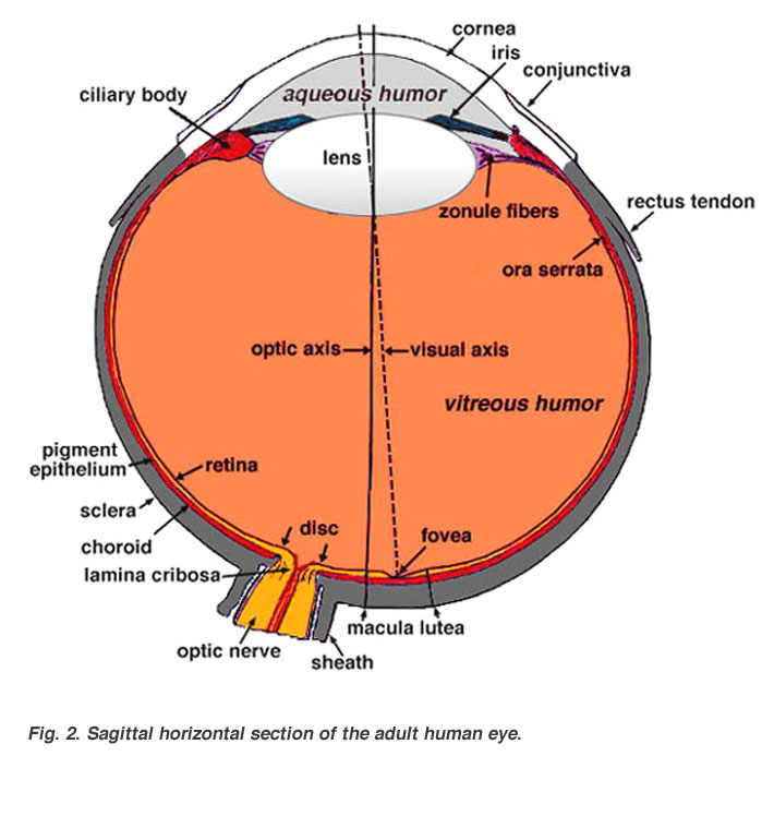
Outer Layer- cornea/sclera
anterior part of sclera covered by mcous menbrane-the conjunctiva
Cornea:
- 3 layers-epithelium, substantia propria(stroma),Descemet's membrane with endothelium
- epithelium-stratified, basal cells lie on Bowman's membrane
- stroma-90% of corneal thickness - regularly arranged thin fibrils of collagen ensheathed by acid mucopolysaccharides set in a ground substance- form ribbon like bundles & give the stroma a laminated appearance- fibrils circular in CS-spaced equidistant- hexagonal lattice
- transparency related to regularity of stromal components-interfibrilar spacing less than a wavelength of light-tangential rows of fibres act as diffraction gratting resulting in destructive interference of scattered rays
- Descemet's membrane-thin elastic membrane-covered on posterior surface by endothelium
- stromal hydration maintained by endothelium- electrolytes removed & water flows passively
- endothelium examined by 500x specular microscope
- endothelial cells decrease in number with age-residual individual cells enlarge to compensate
- corneoscleral junction- limbus- cornea set into a sclera like a watch glass
- nerve supply-trigeminal
- no blood vessels- minute arcades @ limbus (1mm)
- corneal nourishment- diffusion of aqueous humour & peripheral vessels
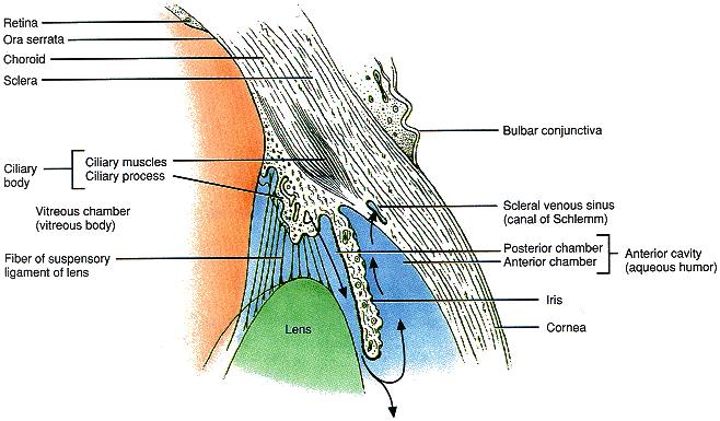
Lining the Inner Aspect of Sclera-
- Uveal Tract-highly vascular-for nutrition
- Retina
Uveal Tract:
- Choroid
- Ciliary Body
- Iris- anteriormost
Anterior Chamber:
- aquous humour
- between cornea & iris 2.5 mm deep in centre
- peripheral recess- angle of anterior chamber
- canal of Schlemm- circular venous sinus in inner layer of sclera- often more than one lumen
- trabecular meshwork between canal of Schlemm & recess of anterior chamber
Iris:
- anterior surface-single layer endothelium-not continuous@crypts
- stroma contains branched connective tissue cells
- iris stroma usually pigmented-unpigmented in blue sclera
- blood vessels in iris-radial
- tissue spaces communicate directly with anterior chamber through crypts@ ciliary border
- thinnest@attachment to ciliary body
- posterior surface-2 pigmented epithelium-developmentally from retina-continuous@pupillary margin-anterior layer flattened cells-posterior layer cuboidal cells
- pupillary smooth muscles derived from from anterior epithelial cells
- sensory- trigeminal
- sphincter pupillae-occulomotor
- dilator pupillae-sympathetic (cervical chain)
Ciliary Body:
- like isosceles triangle- base forwards
- chief mass-ciliary muscle-unstriped-3 parts-circumferential-blends with scleral spur
- most muscles meridional in anteroposterior direction, 2nd portion v-shaped interdigitating concentrically@base of iris, 3rd portion -insertion@ root of iris-just anterior to pigmentary epithelium-closely related to dilator muscle
- anterior surface-corrugated-pars plicata-contains ciliary processes(tufts of blood vessels-like glomeruli) in between
- posterior part-smooth-pars plana
- covered on inner surface by 2 layers of epithelium-belongs to retina-only outer layer pigmented
- posterior extent-ora serrata-transition from ciliary body to choroid gradual
- ora serrata more anterior on nasal side than temporal
- sensory-trigeminal
- motor-occulomotor & sympathetic
Choroid:
- extremely vascular membrane in contact everywhere with sclera with a potential space-epichoroidal/suprachoroidal space in between
- inner side lamina vitrea/membrane of Bruch
- blood vessels increase in calibre from inside to outside-choriocapillaries(fenestrated vessels) immediately beneath membrane of Bruch
- sensory-trigeminal
- autonomic supply-for vasomotor
Retina:
- Outer layer epithelium-hexagonal single layer pigment epithelium
- Inner layer epithelium-suddenly changes@ora serrata into highly complex visual retina
Retina formed by 3 strata of cells & their synapses:
- visual cells-externally
- relay layer of bipolar cells-intermediate
- ganglion cells-internally

- pigment epithelium-hexagonal cells-single layer-assist metabolism of retina- products of metabolism are freely exchanged between receptor cells & pigment epithelium-melanin granules prominent(absorbs light)-phagosomes present-
- rods & cones-neural epithelium-discs renewed continuously-rod discs have limited life,eventually lost to pigment epithelium
- external limiting membrane-perforated by rods & cones
- outer nuclear layer-nuclei of rods & cones
- outer plexiform layer-synaptic layer-transmissive region
- inner nuclear layer-nuclei of bipolar cells-
- inner plexiform layer-synaptic
- ganglion cell layer
- nerve fibre layer-axons of ganglion cells running centrally to optic nerve
- internal limiting membrane-separates retina from vitreous
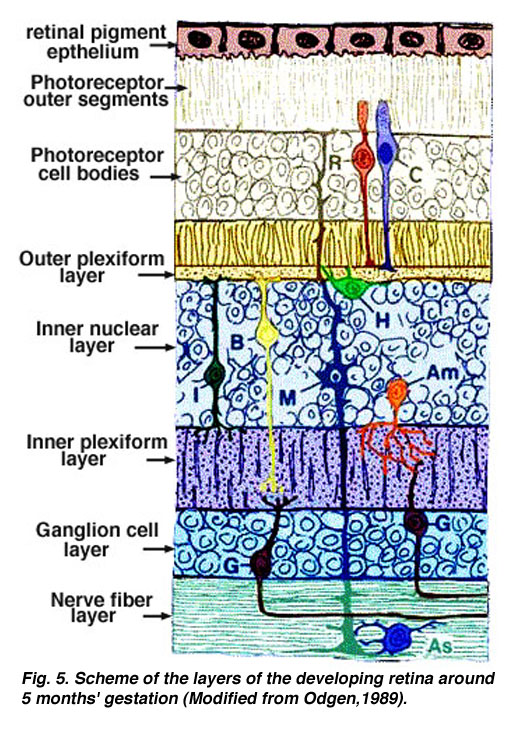
- Fibres of Muller-better developed vertical cells-supportive neuroglial cell- nutritive function
- Fovea Centralis- at posterior pole (3mm in the temporal side of the optic disc)-only cones present- other layers almost completely absent- most sensitive part of retina
- Macula lutea(yellow spot)-surrounds fovea centralis-nuclear layers get thinned out-plexiform layer present-ganglion cells heaped up into several layer(in stead of consisting of a single row of cells)-no blood vessels present-nourishment entirely by choroid)-more sensitive than other parts of retina,less than fovea
- Optic Disc-fibres of nerve fibre layer pass into the optic nerve-other layers of retina stop abruptly@ the edge of the aperture in the scleral canal- spanned by transverse layer of connective tissue containing much elastic tissue(lamina cribrosa)-through its meshes the optic nerve fibres pass-on posterior side the nerve fibres abruptly become surrounded by medullary sheaths
Lens:
Biconcave mass of peculiarly differentiated epithelium-developed from invagination of surface ectoderm of the fetus(compare with plantar corns)-original surface goes inside@centre-peripheral cells correspond to the basal cells of epidermis-inner old cells undergo sclerosis-changes analogous to that of stratum granulosum in epidermis-becomes massed together in the form of nucleus-Lens is devoid of nerve supply
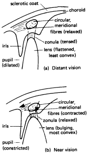
- Accommodation is one of the focusing mechanisms of the eye.Accommodation
- In order to focus rays from objects at varying distances, the lens must change it's refractive power.
- To change it's refractive power, the lens changes shape
- The exact shape of the lens is determined by seventy or so suspensory ligaments.
- The suspensory ligaments attached to the lens are called zonula.
- The zonula are attached radially around the lens.
- The zonula pull the edges of the lens towards the clilary body.
- When the eye is accommodated for distant vision, both the circular and meridional fibres of the ciliary muscle are relaxed. (a)
- When both the circular and meridional fibres of the ciliary muscle are relaxed, the zonula is stretched.
- When the zonula is stretched it pulls the elastic lens into a flattened shape.
- When the eye is accommodated for near vision, both the circular and meridional fibres of the ciliary muscle contract. (b)
- When both the circular and meridional fibres of the ciliary muscle contract, the tension in the zonula is released.
- When the ciliary muscle contracts, the ciliary process and choroid move forward toward the lens.
- When the tension in the zonula is released, it allows the the elastic lens to bulge.
- The lens of the eye is elastic.
- Because the lens is elastic, it bulges, shortens, and thickens.
- The ciliary process is the aqueous humor factory.
- The aqueous humour is drained out of the scleral venous sinus.
No comments:
Post a Comment