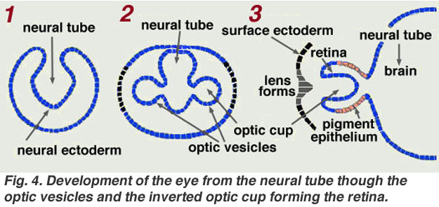
1.Neural Groove- Invaginates to form neural tube, longitudinal, dorsal
2.Optic Plate- thickening from either side of lateral aspect the precursor of forebrain
3. Primary Optic Vesicle-when optic plate grows outwards as a diverticulum towards the surface
4.Optic Cup(secondary optic vesicle)-forms after meeting surface ectoderm, primary optic vesicle invaginates from below
5.Embryonic fissure-the line of invagination during formation of optic cup
6. Inner layer of optic cup- forms main structure of retina-nerve fibres grow from it
7.Outer layer of retina- forms single layer of pigment epithelium
8.The space between retina & pigment epithelium- original primary optic vesicle
9.From anterior border of original optic vesicle develops parts of the ciliary body & iris
10.At the meeting point of neural ectoderm & surface ectoderm, the surface ectoderm thickens to form the lens plate
11.The lens plate invaginates to form lens vesicle & then separates to form lens
12.The hyaloid artery enters the optic cup through embryonic fissure & grows forward to meet the lens, bringing temporary nourishment to the developing structures before it eventually atrophies & disappears-replaced by vitreous-largely secreted by surrounding neural ectoderm
13.The mesoderm surrounding the optic cup differentiates to form the coats of eye & orbital structures, that between the lens & surface ectoderm becomes hollowed to form the anterior chamber, lined by mesodermal condensations which forms the anterior layers of the iris, the angle of anterior chamber & the main structures of cornea.
14.Surface ectoderm remains as corneal & conjunctival epithelium.
15.In the surrounding region, folds grow over in front of the cornea, unite & separate again to to form the lids.
Major Milestones in development of Eye after the Corresponding Periods of Conception:
3wks-Optic groove appears
4th wk-optic pit develops into optic vesicle-lens plate forms-embryonic fissure develops
1month-lens pit & then lens vesicle form-hyaloid vessels develop
11/2 month-closure of embryonic fissure-differentiation of retinal pigment epithelium-proliferation of neural retinal cells-appearance of eyelid folds & nasolacrimal ducts
7th week-formation of embryonic nucleus of lens-sclera begins to form-migration of waves of neural crest(1st wave-corneal & trabecular endothelium,2nd wave-formation of corneal stroma,3rd wave-formation of iris stroma)
3rd month-Differentiation of precursors of rods & cones-anterior chamber appears-fetal nucleus starts developing-sclera condenses-eyelid folds lengthen & fuse
4th month-beginning of formation of retinal vasculature-hyaloid vessel begins to regress-formation of optic disc cup & lamina cribrosa-canal of Schlemm appears-Bowman's membrane develops-formation of major arterial circle & sphincter muscles of iris
5th month-photoreceptors differentiate-eyelid sepration begins
6th month-differentiation of dialator pupillae muscle-nasolacrimal system becomes patent-cones differentiate
7th month-rods differentiate-myelination of optic nerve begins-posterior movement of anterior chamber angle-retinal vessels start reaching nasal peripheri
8th month-completion of anterior chamber angle formation-hyaloid vessels disappear
9th month-retinal vessels reach temporal periphery, pupillary membrane disappears
After Birth-Macular region of the retina develops further
Precursors & Corresponding Derivatives of Eye structures:
Neural Ectoderm- smooth muscle of iris-optic vesicle & cup-iris epithelium-part of vitreous-retina-retinal pigment epithelium,fibres of optic nerve
Surface Ectoderm-conjunctival epithelium,corneal epithelium,lacrimal glands,tarsal glands & lens
Mesoderm-extraocular muscles,corneal stroma,sclera,iris,vascular endothelium of eye & orbit,choroid, part of vitreous
Neural Crest-corneal stroma,keratocytes & endothelium; sclera; trabecular meshwork endothelium, iris stroma, ciliary muscles, choroidal stroma, part of vitreous, uveal & conjunctival melanocytes, meningeal sheaths of optic nerve,ciliary ganglion,Schwann cells of ciliary nerves,orbital bones,orbital connective tissue,connective tissue sheath & muscular layer of ocular & orbital vessels

No comments:
Post a Comment